About Us
The labs current research focus areas include:
Imaging and Solid Modeling
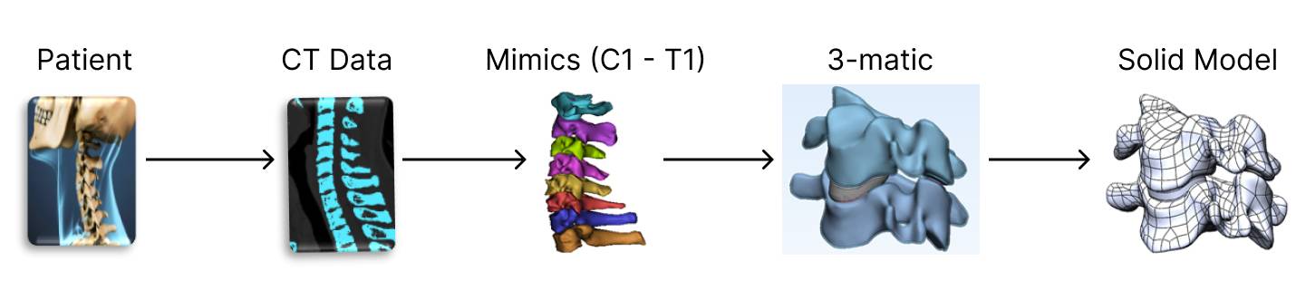 Based on patient’s MRI or CT scan anatomical images and Mimics software, the platform will rapidly create a 3D solid model of patient’s spine using the AI reconstruction system supported by an optimized physiological anatomical-based 3D convolutional neural network that can automatically achieve the computed tomography angiography reconstruction of spine.
Finite Element Biomechanics Simulation
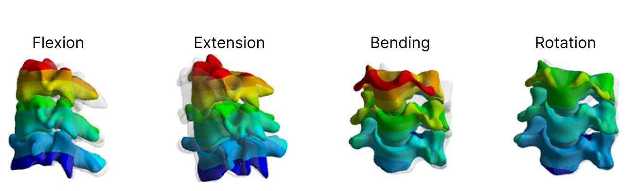
Finite element software such as Ansys and Abaqus will be used for the simulation. The typical process to build a validated, patient-specific finite element model of spine involves: (a) 3D solid model obtained from the MRI or CT scan, and the implant design obtained from 3D printer; (b) material properties and constitutive model for the implant and each spine components such as vertebrae, ligaments, intervertebral disc, facet joints and muscles, etc.; (c) definition of boundary conditions, loads, constraints, and contact conditions; (d) mesh generation, convergent test, validation and sensitivity analysis; (e) develop dependable model of cadaver specimens to establish the error bounds for patient-specific model validation.
Spinal Implants Design/Analysis and 3D printing
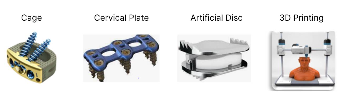
Finite element software such as Ansys and Abaqus will be used for the simulation. The typical process to build a validated, patient-specific finite element model of spine involves: (a) 3D solid model obtained from the MRI or CT scan, and the implant design obtained from 3D printer; (b) material properties and constitutive model for the implant and each spine components such as vertebrae, ligaments, intervertebral disc, facet joints and muscles, etc.; (c) definition of boundary conditions, loads, constraints, and contact conditions; (d) mesh generation, convergent test, validation and sensitivity analysis; (e) develop dependable model of cadaver specimens to establish the error bounds for patient-specific model validation. A 3D printed Cadaver will be developed and sensitized with various developed biosensors. The cadaver model will be validated using values of literature and the intact model of the same spine. The biosensors will be validated using the FEA model.
Patient Spine Monitoring App
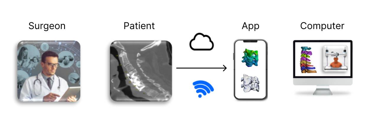
Micro biosensors will be developed and implanted in the cadaver to measure the stress/force and range of motion in the spine, and the subsidence and micromotion of the implants such as cage and artificial disc. The produced and tested biosensors will be implanted into cervical instrumentation for retrieval of in vivo information that will be sent to an app accessed by the patients and clinical team for post-operative follow up. The data will also be uploaded to a cloud data base that will serve as the standard of validation for future developed FEA. These FEA will then be used to improve the next generation of biosensors and so on until both simulation methodologies and biosensing capabilities are perfected.